Click to WhatsApp
 +91-9899993637
 It is a new minimally invasive technique that involves the introduction of image guided electric needles through small punctures in the skin of the body overlying the diseased tissue. The probes are guided to the target site with the help of CT scan or ultrasound. Once the target is reached high frequency current is passed through the needle probes which produces large amount of heat within and around the tumor cells and kills them tumor. It is helpful in localized tumors and can also be used in conjunction with resection or chemotherapy.
RFA can be used successfully to destroy tumor tissue in any part of the body. It can also be used to treat any dysfunctional tissue. Currently it has been used effectively to treat cardiac arrhythmia, liver cancer, varicose veins, kidney tumors, chronic low back pain (rhizotomy). In the last 15 years RFA has come to be a dependable and safe procedure with promising outcome. It is generally used for multiple tumors within the same organ or various organs, primary or secondary tumors, or if the tumor is too large to be resected or if the patient cannot tolerate surgery.
An important advantage of RF current (over previously used low frequency AC or pulses of DC) is that it does not directly stimulate nerves or heart muscle and can therefore often be used without the need for general anesthetic. RFA has become increasingly accepted in the last 15 years with promising results.[1][2] RFA procedures are performed under image guidance (such as X-ray screening, CT scan or ultrasound) by an interventional pain specialist (such as an anesthesiologist), interventional radiologist or a cardiac electrophysiologist, a subspecialty of cardiologists.
Radiofrequency ablation (RFA) Procedure:
 The procedure is performed with patient under conscious sedation (a state of consciousness achieved through medication where the patient can bear unpleasant procedure while maintaining cardiorespiratory function. It is now called as procedural sedation) using drugs like midazolam, fentanyl and meperidine. These drugs are introduced into the body intravenously. The instruments that are used for this procedure include image guiding technique like ultrasound, MRI or CT scan, a radiofrequency generator to produce radiofrequency waves, straight needles and straight hollow needles with retractable electrodes, insulating wires and earthing pads, and an endoscope in case of a laparoscopic procedure is done. The ablation procedure is performed by a specifically trained interventional radiologist in an operation theatre or a radiology room.
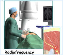 The patient is connected to a monitor where his pulse, respiratory rate and blood pressure are monitored continuously throughout the procedure. In case the patient is sedated under general anesthesia, a tube is inserted down the wind pipe and connected externally to an artificial breathing unit (ventilator). The area where the needles are to be inserted is sterilized and draped. The grounding pads are attached to the back or thigh of the patient. The needles can be introduced into the patient’s body surgically, percutaneously or through endoscope. In the percutaneous method the surgeon makes small punctures in the skin of the abdomen through which the needle probes are inserted. The probe is guided towards the site of tumor using the image guiding option of US, MRI or CT scan or microscopic camera. Once the needle is inside the target tissue, radiofrequency energy is generated by the generator that passes between the needle probes and grounding pads.
This creates a large amount of ionic vibration around the needle which in turn produces a lot of heat. This large amount of heat destroys the cancer (or diseased cells) cells and closes the nearby feeding blood vessels, ensuring no nutrition to the tumor cells. The dead cells shrink away and are replaced by scar tissue. If the tumor is large or located at multiple places in the same organ, the needles are removed and replaced into the various tumor sites for complete destruction of the cancer. This helps to prevent recurrence to a large extent. After the procedure the needles are pulled out and pressure is put at the entry site to prevent any bleeding. The skin is covered with a simple dressing. No sutures are required.
To know more about cancer, Kidney and Lung Tumors in India, please visit this link : https://safemedtrip.com/medical-services/cancer-treatment-in-india.html