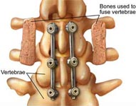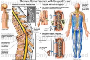
In case the above treatments do not work, the best long-term outcome could be achieved with a Lumbar Fusion.
Doctors at SafeMedTrip.com affiliated Hospitals in India have expertise and vast experience in performing Spinal Fusion – an Advanced minimally invasive surgery is designed to stop the motion at a painful vertebral segment, which in turn should decrease pain generated from the joint. A severely degenerated disc between two vertebral bones or to a “slippage” of the spine bones (which is referred to as “spondylolisthesis”). Slippage of the bones results in misalignment of the spine and possible entrapment of the spinal nerves.

The procedure is performed in an advanced surgical center with Intra-operative MRI guidance. The patient is monitored with an electrocardiogram, blood pressure cuff and blood oxygen-monitoring device.
Medical evaluation includes a physical exam. Diagnostic tests such as MRI (magnetic resonance imaging), steroid injection or discography may be conducted to diagnose and locate the symptomatic disc herniation and determine if the procedure is appropriate.
An incision may be made on abdominal wall, flank, backside, or a combination of these approaches. “Anterior” means “from the front” and “posterior” means from your backside. titanium metal screws would be beneficial. These are typically placed through the “pedicle” bone of the spine and thereby allow purchase of the back, middle, and front parts of your vertebral bones. These screws are attached to a rod or plate that is contoured to your spine. This type of spinal “instrumentation” provides immediate strength to the spine during the process of bone fusion.
In other situations, a titanium metal “cage” or bone cylinder may be screwed into your disc space. This is called an interbody fusion because it is between the “bodies” of the vertebral bones and is across the diseased disc space. This can be done from the front (anterior) or from the backside (posterior). An approach through the abdomen (anterior) may be done “laparoscopically” with several small incisions or “open” through a single incision.
To know more about Spinal Fusion Surgery in India please visit this link : https://safemedtrip.com/medical-services/spine-surgery-treatment-in-india/minimally-invasive-spinal-fusion-surgery-in-india.html

 Click to WhatsApp
 +91-9899993637


