There are many kinds of heart defects. Some are minor, and others are more serious. Defects can occur inside the heart or in the large blood vessels outside the heart. Some heart defects may need surgery right after the baby is born. For others, your child may be able to safely wait for months or years to have surgery.

Paediatric surgeries deal with the correction of abnormalities present in the heart of new-borns, infants or children. Most of the defects are present since birth (congenital). They all come under the category of congenital heart defects. Some are present since birth but cause symptoms after a few years and some cause symptoms immediately after birth.

The major cardiac defects include
- Atrial Septal Defect
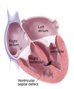
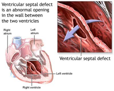
- Ventricular Septal Defect,
- Fallot’s Tetralogy
- Valvular defects
One surgery may be enough to repair the heart defect, but sometimes a series of procedures is needed. Three different techniques for fixing congenital defects of the heart in children are described below. Open-heart surgery is when the surgeon uses a heart-lung bypass machine.
- An incision is made through the breastbone (sternum) while the child is under general anesthesia (the child is unconscious and does not feel pain).
- Tubes are used to re-route the blood through a special pump called a heart-lung bypass machine. This machine adds oxygen to the blood and keeps the blood warm and moving through the rest of the body while the surgeon is repairing the heart.
- Using the machine allows the heart to be stopped. Stopping the heart makes it possible to repair the heart muscle itself, the heart valves, or the blood vessels outside the heart. After the repair is done, the machine is removed, and the heart is started again. The breastbone and the skin incision are closed up.
For some heart defect repairs, the incision is made on the side of the chest, between the ribs. This is called a thoracotomy. It is sometimes called closed-heart surgery. This surgery is done using special instruments and a camera. Another way to fix defects in the heart is to insert a few small tubes into an artery in the leg and pass them up to the heart. Only some heart defects can be repaired this way.
ASD- Atrial Septal Defect -ASD- Atrial Septal Defect refers to a hole in the septum that separates the right and left atrium. This results in mixing of pure and impure blood. Depending on the size of the defect, the symptoms may range from no symptoms to bluish discoloration of the body, increase pulmonary blood pressure and irregular cardiac contractions (arrhythmias). It is corrected by open heart surgery. The sternum is split in the midline. Arterial and double venous (superior vena cava and inferior vena cava) vessels are clamped. By applying cardiopulmonary bypass (heart lung machine), the aorta is clamped, and the heart is temporarily paralyzed by a solution. The right atrium is opened and the defect is sutured.

Two types of surgeries are available –
- Intra-Cardiac Technique – the patient is attached to an external heart-lung machine (which performs circulatory and respiratory function during surgery). It is an open heart surgery in which the patient is placed under general anaesthesia. The rib cage is cut open in the midline. The heart is directly accessed and the defect is sutured.
- Trans- Catheter Technique – Surgical instruments are passed through the catheter which is inserted in the femoral artery at the groin. The catheter is slowly guided up towards the point of defect to close it. It is a successful surgery. With the separation of ventricles normal circulation resumes. The enlarged heart comes back to normal size. Pulmonary artery pressure reduces. The child’s growth fastens. The prognosis is excellent.
Double valve repair and replacement
| Valves are openings between two chambers and also between a chamber and artery which allows unidirectional flow of blood. The patency of these valves is very important for normal flow of blood. The function of mitral valve (between left atrium and left ventricle) and the aortic valve (between left ventricle and aorta) is very important. If these valves get narrowed, the amount of blood passing from left atrium to left ventricle (mitral valve) or from left ventricle to aorta (aortic valve) is drastically reduced. In this case the heart will pump blood harder to push it through the narrowed valves. But the blood will tend to go upwards back into the pulmonary vein and finally to the lungs. This will cause excess blood in the lungs leading to congestion. At the same time very less blood will come out into the aorta from the left ventricle which will result in reduced blood and oxygen supply to all the body parts. |
To know more about “Pediatric Cardiac Surgery” in India, Please visit this link please check out this page https://safemedtrip.com/medical-services/heart-surgery-in-india/pediatric-cardiac-surgery-india.html

 Click to WhatsApp
 +91-9899993637


