The high incidence of lung cancer and the poor survival rate make lung cancer a very important public health problem, and the leading cause of cancer death in the world. Most patients have locally advanced lung cancer at the time of diagnosis. As per the Madras Metropolitan Tumour Registry (MMTR) Chennai, in 2006-2008, cancer of the lung was the most common among males, and was ranked among the top ten in females.
It constituted 10.9 percent and 3.3 per cent of all cancers among males and females respectively. The peak incidence occurred in the age group of 65-69 years among both sexes.
Risk factors:
Cigarette smoking is the single highest cause of the lung cancer epidemic.
Other contributing factors are asbestos, arsenic, chromium, nickel, and radon in the work environment.
Other environmental factors such as passive smoking and air pollution.
Molecular changes that commonly occur in lung cancer are mutations of tumour suppressor genes p16, p53, and H, K, N-ras family of oncogenes.
Studies have shown that individuals with a higher dietary intake of fruits or vegetables have a lower risk of lung cancer.
Types
Lung cancers are classified into:
- Non-Small Cell Carcinoma (NSCLC) that includes squamous cell carcinoma, adenocarcinoma, large cell carcinoma and their subtypes (80 per cent).
- Small Cell Carcinoma (SCLC) (20 percent).
- There are other rare types of lung cancers that can occur apart from these.
Prognostics
Well differentiated squamous cell carcinomas and non mucinous bronchiloalveolar carcinomas have favourable prognosis.
Poor prognostic factors include: higher tumour size and extent, regional nodal involvement, absence or presence of distant metastasis, weight loss of more than 10 per cent, age less than 40 years, tumour size more than 3 cm, Iymphovascular invasion, and mutation of the tumour suppressor gene p53.
Spread
Spread can occur along bronchus into lung parenchyma, to mediastinum or pleura causing pleural effusion. Diaphragm and chest wall involvement are not uncommon. 50 per cent have nodal metastasis at resection. Distant spread commonly involves adrenals to 50 per cent, liver to 30 percent, apart from brain, bone opposite lung, pericardium and kidneys.
Common Symptoms
Cough, weight loss, chest pain, shortness of breath, blood in the sputum, superior vena cava syndrome, ulnar nerve and Horner’s syndrome (Pancoast tumour) are common symptoms of lung cancer.
Screening for lung cancer

At present, screening for early detection of lung cancer is not recommended, probably because of the failure of early studies to demonstrate any mortality reduction from lung cancer evaluation based on sputum cytology and/or chest radiography. With the introduction of helical computerised tomography, a new imaging modality that can detect nodules as small as a few millimetres, the potential benefits of lung cancer screening is being re-examined.
Imaging in lung cancer
Chest radiography remains the basic modality for the detection of lung cancer.
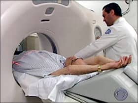
Computerised tomography (CT) provides information about the primary lesion, thoracic lymph nodes, pleura, chestwall and upper abdomen. It is the standard imaging modality for staging lung cancer.
Magnetic resonance imaging (MRI) appears to be superior to CT in detecting mediastinal, chest wall tumour invasion into the pericardium, heart and great vessels, brachial plexus, vertebral body and spinal canal.
Positron emission tomography (PET) is a molecular imaging modality that detects metabolic changes in tumour cells. PET improves the rate of detection of the extent of primary tumour, draining nodes and distant metastases thereby improving the staging accuracy in patients with NSCLC that can have a significant impact on clinical management.
Other diagnostic modalities
Clinical and radiological findings should guide the diagnostic approach, depending on the size and location of the tumour, the presence of metastatic disease, and the clinical status of the patient. Diagnostic and staging work is taken up concomitantly.
Sputum cytology, flexible bronchoscopy for biopsies, brushings and washings, CT guided transthoracic needle aspiration, oesophageal endoscopic ultrasound guided fine needle aspiration/trucut biopsy of the mediastinal nodes, anterior mediastinoscopy to assess lymph nodes, are the aids used to establish the histopathological diagnosis. Distant metastatic sites need to be documented with microscopic diagnosis.
Management
- Surgery is the preferred modality of primary management for resectable NSCLC.
- Based on initial stage and postoperative histopathological report, patients will be planned for adjuvant radiation therapy with or without chemotherapy, or chemotherapy with or without radiation therapy.
- For medically inoperable and unresectable tumours chemoradiation therapy is the preferred line of management.
- With the rapid technological explosion in diagnostic imaging and radiation delivery techniques, radiation oncologists are now able to deliver external beam radiation therapy with high precision using Image Guided Intensity Modulated Radiation Therapy (IG IMRT), Stereotactic Body , Radio surge with real time positional management respiratory gating system delivering significantly higher doses to the tumour and minimum dose to the surrounding normal lung and other critical structures like the opposite lung, heart, spinal cord, oesophagus, and breast, resulting in increased cure rates and lesser side effects respectively.
- Radiation along with chemotherapy has a role in palliation of symptoms due to recurrent, advancing and metastatic cancer.
- Newer targeted therapy compounds have resulted in progression-free and overall survival advantage in NSCLC.
- For SCLC, chemoradiation therapy is the preferred choice of treatment, except in T1-2 NO MO where surgery followed by chemotherapy is the standard of care.
The by and large outcome for patients with lung cancer may not be gratifying at present. But with improved surgical techniques, newer anti-cancer drugs, and modern radiation therapy delivery techniques, we are able to confer superior progression-free and overall survival period ensuring a good quality of life for our patients. Lung cancer surgery hospitals in India attracts many international patients because of availability of very good treatment facilities, good success rate and very low cost of treatment.
Get Free Expert Opinion Online from India’s Most Experienced Lung Cancer Specialists Right Now
Get FREE Cervical Cancer Treatment Cost Estimates from Top Hospitals in India.
We help you get FREE Expert Opinion from Top Doctors at Best Hospitals in All Major Cities in India.
We understand each patient is unique and therefore, we offer customised treatment packages that include medical visa assistance for international patients, assistance in certified hospital/doctor selection, finalising your appointments, etc and that too inclusive of room rent, consultation fees, investigation charges, nursing, diet and drugs.
So, no matter how complex is the surgery, we will help you choose a doctor considering your requirement, budget and availability.
Since our inception in 2010, we have helped many patients lead a normal and quality life. We assist you in choosing the best hospital in India, seeking an appointment with a qualified and experienced cancer specialist, explore your options, assist in a medical tourist visa, besides offering other services such as airport transfers, appoint an interpreter (if need be), etc.
If you have any further queries or in case you need any assistance, speak to the Case Manager or log on to our website at www.safemedtrip.com

 Click to WhatsApp
 +91-9899993637


